Biomedical Optics & Medical ImagingSimulation techniques enhance cellular nanobioimagingStoyan Tanev, James Pond, Paul Paddon, and Valery TuchinA new 3D computational approach constructs realistic optical phase contrast microscope images of gold nanoparticles in biological cells.
Biomedical optics researchers need optical simulation tools to acquire a deeper understanding of the interactions between light and tissues.1,2 The challenges associated with the modeling of light scattering from single cells come from two major factors. First, the wavelength of light is comparable to the size of the scattering sub-cellular structures. Second, biological cells have irregular shapes and arbitrarily distributed refractive indices, which makes it impossible to use analytical modeling approaches. Both factors necessitate the use of numerical simulation methods based on rigorous electromagnetic theory. These include: the method of separation of variables, the finite element method, the method of lines, the point matching method, the method of moments, the discrete dipole approximation method, the null-field (extended boundary condition) method, the T-matrix electromagnetic scattering approach, the surface Green's function electromagnetic scattering approach, and the finite-difference time-domain (FDTD) method.3
Figure 1. Schematic representation of the FDTD computational domain. NPs: nanoparticles.
Figure 2. a) Schematic representation of an optical phase contrast microscope (OPCM). b) 2D visual representation of the FDTD OPCM model.The two main advantages of the FDTD method are: its numerical simplicity and straightforward physical basis (it is a numerical expression and solution of Maxwell's equations); and its ability to be easily integrated with a graphical user interface, which enables its broader adoption as a biomedical research tool. Its main disadvantages are: first, its computer power and memory requirements, and second, the need of more sophisticated post-processing techniques. The first of the disadvantages is becoming less of an issue due to the growing computational capabilities and the affordability of powerful commercially available computers and grid computing resources. Our research program is addressing many of the aspects associated with the second disadvantage.The FDTD method4–10 gained significant popularity in studying the light scattering patterns5,7–9 from single cells including the effect optical immersion11–13 but did not provide yet a way to the modeling of cell imaging. We developed a new way of using the 3D FDTD approach to construct optical phase contrast microscope (OPCM) images of cells containing gold nanoparticle (GNP) clusters.8The FDTD technique is a numerical solution of Maxwell's equations describing the properties of an electromagnetic field relative to the source, charge density, and current density.14 FTDT methods normally use an input plane to propagate a linearly polarized wave in a finite region containing the cell (see Figure 1).14 The total near fields are recorded at a monitoring plane behind the cell and then projected to the far field using the FTDT near-to-far-field algorithm.14,15The OPCM images are created by the interference of an incident reference (R) and a diffracted (D) beam–see Figure 2(a). The incoherent annular illumination of the OPCM is modeled by adding up the results of eight different simulations, as shown in Figure 2(b). These are generated under two polarizations using input plane waves incident at a given polar angle of 30° or an azimuthal angle of either 0°, 90°, 180° or 270°. The optical lens system is characterized by a magnification factor of 10 and a numerical aperture of 0.8.We modeled a cell with a cluster of 42 GNPs using the radius of the nucleus and of the entire cell and the refractive indices of the cytoplasm, nucleus, cell membrane, and extracellular environment (see Figure 3). We then constructed OPCM images of the cell at the refractive index matching (RIM) condition: ncytoplasm=nextra−cell=1.36 for a wavelengths of 543.0nm (GNP resonance) and 676.4nm (no GNP resonance) (see Figure 4).13,16–21 The enhanced GNP cluster imaging at resonant conditions is clearly visible in Figure 5.
Figure 3. The cell model's size (in radius, R, or thickness, d) and index of refraction (n) characteristics: Rcell=5μm, ncytoplasm=1.36, nextra-cell=1.33), 10nm < dmembrane < 20nm, nmembrane=1.47, Rnucleus=1.5μm, nnucleus=1.40. The cell contains a cluster of 42 gold NPs (GNPs) with Rnanoparticle=50nm.
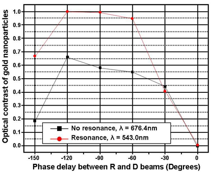
Figure 4. OPCM cell images for phase offset ψ=- 120°. The GNP cluster is to the right of the nucleus as in the model in Figure 3.http://spie.org/Images/Graphics/Newsroom/Imported/1224/1224_fig5b.jpgFigure 5. Maximum optical contrast of the GNP cluster as a function of the phase offset Ψ.Our results demonstrate the capability of the proposed FDTD OPCM approach to model enhanced cell imaging due to GNPs. The method could provide insights for the application of GNPs in optical nanotherapeutics. We are currently working on extending our model to include the relationship between the optical and thermal properties of gold nanospheres, nanoshells, nanorods, and nanosphere clusters, which is crucial for the application of phototherapeutic effects for the selective treatment of cancer cells, bacteria, viruses, and DNA.We would like to acknowledge the use of computing resources from WestGrid. This work was supported by grants from the Federal Agency of Education of RF No 1.4.06, RNP.2.1.1.4473, RFBR No. 06-02- 16740, and Photonics4Life of FP7. We all thank Dr. V. Zharov for fruitful discussions.Stoyan TanevCarleton UniversityOttawa, Canadahttp://www.sce.carleton.ca/faculty/tanevhttp://www.sce.carleton.caStoyan Tanev received his MSc and PhD in Physics in 1995 from the University of Sofia, Bulgaria, and the University Pierre and Marie Curie, Paris, France. He received a MEng in Technology Management in 2005 from Carleton University. His research interests are in nanobiophotonics design and modeling, and in the application of open innovation principles in emerging technology areas.James Pond, Paul PaddonLumerical Solutions Inc.Vancouver, CanadaValery TuchinInstitute of Optics and BiophotonicsSaratov State UniversitySaratov, RussiaInstitute of Precise Mechanics and ControlRussian Academy of SciencesSaratov, RussiaReferences:1. P. Prasad, Introduction to Biophotonics, ch. 7, John Wiley & Sons, New Jersey, 2003.2. V. Tuchin, Tissue Optics: Light Scattering Methods and Instruments for Medical Diagnosis, SPIE Publications, 2007.3. F. M. Kahnert, Numerical methods in electromagnetic scattering theory, J. Quantitative Spectroscopy and Radiative Transfer 73, pp. 775-824, 2003.4. F. Kahnert, Numerical methods in electromagnetic scattering theory, J. Quant. Spectrosc. Radiat. Transfer 79-80, pp. 775, 2003.5. R. Drezek, A. Dunn, R. Richards-Kortum, A pulsed finite-difference time-domain (FDTD) method for calculating light scattering from biological cells over broad wavelength ranges, Optics Express 6, pp. 147, 2000.6. T. Tanifuji, M. Hijikata, Finite difference time domain (FDTD) analysis of optical pulse responses in biological tissues for spectroscopic diffused optical tomography, IEEE Trans. Med. Imag. 21, pp. 181, 2002.7. R. Drezek, M. Guillaud, T. Collier, I. Boiko, A. Malpica, C. Macaulay, M. Follen, R. Richards-Kortum, Light scattering from cervical cells throughout neoplastic progression: influence of nuclear morphology, DNA content, and chromatin texture, J Biomed. Opt. 8, pp. 7, 2003.8. S. Tanev, V. Tuchin, P. Paddon, Cell membrane and gold nanoparticles effects on optical immersion experiments with noncancerous and cancerous cells: finite-difference time-domain modeling, J. Biomed. Opt. 11, pp. 064037, 2006.9. X. Li, A. Taflove, V. Backman, Recent progress in exact and reduced-order modeling of light-scattering properties of complex structures, IEEE J. Selected Topics Quant. Electron. 11, pp. 759, 2005.10. R. Barer, K. Ross, S. Tkaczyk, Refractometry of living cells, Nature 171, pp. 720, 1953.11. B. A. Fikhman, Microbiological Refractometry, Medicine, Moscow, 1967. (in Russian)12. V. Tuchin, Optical Clearing of Tissues and Blood, SPIE Publications, 2005.13. S. Tanev, V. Tuchin, P. Paddon, Light scattering effects of gold nanoparticles in cells: FDTD modeling, Laser Phys. Lett 3, pp. 594, 2006.14. A. Taflove, S. Hagness, Computational Electrodynamics: The Finite-Difference Time Domain Method, Artech House Publishers, 2005.15. The simulations were performed by the FDTD Solutions trademark software. http://www.lumerical.com16. K. Sokolov, M. Follen, J. Aaron, I. Pavlova, A. Malpica, R. Lotan, R. Richards-Kortum, Real-time vital optical imaging of precancer using anti-epidermal growth factor receptor antibodies conjugated to gold nanoparticles, Cancer Research 63, pp. 1999-2004, 2003.17. I. El-Sayed, X. Huang, M. El-Sayed, Surface plasmon resonance scattering and absorption of anti-EGFR antibody conjugated gold nanoparticles in cancer diagnostics: applications in oral cancer, Nano Lett. 5, pp. 829, 2005.18. N. Khlebtsov, A. Melnikov, L. Dykman, V. Bogatyrev, Optical properties and biomedical applications of nanostructures based on gold and silver bioconjugates, Photopolarimetry in Remote Sensing, pp. 265, Kluwer, Dordrecht, 2004.19. V. Zharov, J. Kim, D. Curiel, M. Everts, Self-assembling nanoclusters in living systems: application for integrated photothermal nanodiagnostics and nanotherapy, Nanomedicine: Nanotechnology, Biology, and Medicine 1, pp. 326, 2005.20. V. Zharov, K. Mercer, E. Galitovskaya, M. Smeltzer, Photothermal nanotherapeutics and nanodiagnostics for selective killing of bacteria targeted with gold nanoparticles, Biophys. J. 90, pp. 619, 2006.21. P. Johnson, R. Christy, Optical constants of the noble metals, Phys. Rev. Lett. B 6, pp. 4370, 1972.DOI: 10.1117/2.1200808.1224


Comments