Traz Ardagau's Posts (11)

Sort by

Recent use of fluorescent labels in nanotechnology drug delivery, medical, biochemical and biological science:


http://www.lumiprobe.com/citations

Nooney, R.; O’Connell, C.; Roy, S.; Boland, K.; Keegan, G.; Kelleher, S.; Daniels, S.; McDonagh, C. Synthesis and characterisation of far-red fluorescent cyanine dye doped silica nanoparticles using a modified microemulsion method for application in bioassays. Sensors and Actuators B: Chemical, 2015, in press. doi:  10.1016/j.snb.2015.06.117

Li, H.; Yang, Z.-Y.; Liu, C.; Zeng, Y.-P.; Hao, Y.-H. Gu, Y.; Wang, W.-D.; Li, R. PEGylated ceria nanoparticles used for radioprotection on Human liver cells under γ-ray irradiation , 2015, in press. doi: 10.1016/j.freeradbiomed.2015.06.010
 
 Ma, Y.; Fuchs, A.; Boase, N.R.B.; Rolfe, B.E.; Coombes, A.G.A.; Thurecht, K.J. The in vivo fate of nanoparticles and nanoparticle-loaded microcapsules after oral administration in mice: evaluation of their potential for colon-specific delivery. European Journal of Pharmaceutics and Biopharmaceutics, 2015, 94, 393–403. doi:   10.1016/j.ejpb.2015.06.014


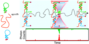
Noriega, R.; Finley, D.T.; Haberstroh, J.; Geissler, P.L.; Francis, M.B.; Ginsberg, N.S. Manipulating Excited-State Dynamics of Individual Light-Harvesting Chromophores through Restricted Motions in a Hydrated Nanoscale Protein Cavity. The Journal of Physical Chemistry B, 2015, 119(23), 6963–6973. doi: 

10.1021/acs.jpcb.5b03784 


 Shi, Y.; van der Meel, R.; Theek, B.; Oude Blenke, E.; Pieters, E.H.E.; Fens, M.H.A.M.; Ehling, J.; Schiffelers, R.M.; Storm, G.; van Nostrum, C.F.; Lammers, T.; Hennink, W.E. Complete Regression of Xenograft Tumors upon Targeted Delivery of Paclitaxel via Π–Π Stacking Stabilized Polymeric Micelles. ACS Nano, 2015, 9(4), 3740–3752. doi: 10.1021/acsnano.5b00929

Duong, H.T.T.; Dong, Z.; Su, L; Boyer, C.; George, J.; Davis, T.P.; Wang, J. The Use of Nanoparticles to Deliver Nitric Oxide to Hepatic Stellate Cells for Treating Liver Fibrosis and Portal Hypertension. Small, 2015, 11(19), 2291–2304. doi:  10.1002/smll.201402870 


 Wen, A.M.; Infusino, M.; De Luca, A.; Kernan, D.L.; Czapar, A.E.; Strangi, G.; Steinmetz, N.F. Interface of Physics and Biology: Engineering Virus-Based Nanoparticles for Biophotonics. Bioconjugate Chemistry, 2015, 26(1), 51–62. doi:     10.1021/bc500524f

Nooney, R.I.; White, A.; O’Mahony, C.; O’Connell, C.; Kelleher, S.M.; Daniels, S.; McDonagh, C. Investigating the colloidal stability of fluorescent silica nanoparticles under isotonic conditions for biomedical applications. Journal of Colloid and Interface Science, 2015, 456, 50–58. doi:   10.1016/j.jcis.2015.05.051


Lumiprobe citation list
- research publications citing use of fluorescent reagents, sortable by date or cited product.
- cyanine NHS esters, amines, azides, alkynes, hydrazides, maleimides, carboxylic acids


Lumiprobe catalog 


Other citations:
http://www.lumiprobe.com/citations

Protocols
http://www.lumiprobe.com/protocols

Lumiprobe sponsors nanpaprika.eu
For 5% discount when you order use code:   nanopap

Read more…

Lumiprobe offers fluorescent labels dyes, click chemistry reagents

Recent use of fluorescent labels in nanotechnology drug delivery, medical, biochemical and biological science:

Li, L.; Sun, W.; Zhong, J.; Yang, Qi.; Zhu, X.; Zhou, Z.; Zhang Z.; Huang, Y. Multistage Nanovehicle Delivery System Based on Stepwise Size Reduction and Charge Reversal for Programmed Nuclear Targeting of Systemically Administered Anticancer Drugs. Advanced Functional Materials, 2015. doi: 10.1002/adfm.201501248

Wilks, M.Q.; Normandin, M.D.; Yuan, H.; Cho, H.; Guo, Y.; Herisson, F.; Ayata, C.; Wooten, D.W.; El Fakhri, G.; Josephson, L. Imaging PEG-Like Nanoprobes in Tumor, Transient Ischemia, and Inflammatory Disease Models. Bioconjugate Chemistry, 2015, in press. doi: 10.1021/acs.bioconjchem.5b00213

Abolmaali, S.; Tamaddon, A.; Kamali-Sarvestani, E.; Ashraf, M.; Dinarvand, R. Stealth Nanogels of Histinylated Poly Ethyleneimine for Sustained Delivery of Methotrexate in Collagen-Induced Arthritis Model. Pharmaceutical Research, 2015, in press. doi: 10.1007/s11095-015-1708-0

Zhang, J.; Wang, G.; Mao, D.; Han, A.; Xiao, N.; Qi, X.; Ding, D.; Kong, D. Targeted In Vivo Imaging of Mouse Hindlimb Ischemia Using Fluorescent Gelatin Nanoparticles. Journal of Nanomaterials, 2015, 2015, Article ID 704817

Unciti-Broceta, J.D.; Cano-Cortés, V.; Altea-Manzano, P.; Pernagallo, S.; Díaz-Mochón, J.J.; Sánchez-Martín, R.M. Number of Nanoparticles per Cell through a Spectrophotometric Method – A key parameter to Assess Nanoparticle-based Cellular Assays. Scientific Reports, 2015, 5, 10091. doi: 10.1038/srep10091

Choi, E.B.; Choi, J.; Bae, S.R.; Kim, H.-O.; Jang, E.; Kang, B.; Kim, M.-H.; Kim, B.; Suh, J.-S.; Lee, K. et al. Colourimetric redox-polyaniline nanoindicator for in situ vesicular trafficking of intracellular transport. Nano Research, 2015, 8(4), 1169–1179. doi: 10.1007/s12274-014-0597-6

Hsueh, P.-Y.; Edman, M.C.; Sun, G.; Shi, P.; Xu, S.; Lin, Y.-a.; Cui, H.; Hamm-Alvarez, S.F.; MacKay, J.A. Tear-mediated delivery of nanoparticles through transcytosis of the lacrimal gland. Journal of Controlled Release, 2015, 208, 2–13. doi: 10.1016/j.jconrel.2014.12.017

Hou, Z.; Lin, J.; Li, Y.; Guo, F.; Yu, F.; Wu, H.; Fan, Z.; Zhi, L.; Luo, F. Validation of a dual role of methotrexate-based chitosan nanoparticles in vivo. RSC Advances, 2015, 5(52), 41393–41400. doi: 10.1039/c5ra03705K

Kim, M.-G.; Park, J.Y.; Miao, W.; Lee, J.; Oh, Y.-K. Polyaptamer DNA nanothread-anchored, reduced graphene oxide nanosheets for targeted delivery. Biomaterials, 2015, 48, 129–136. doi: 10.1016/j.biomaterials.2015.01.009

Nakamura, T.; Sugihara, F.; Matsushita, H.; Yoshioka, Y.; Mizukami, S.; Kikuchi, K. Mesoporous silica nanoparticles for 19F magnetic resonance imaging, fluorescence imaging, and drug delivery. Chemical Science, 2015, 6, 1986–1990. doi: 10.1039/c4sc03549f


Other citations:
http://www.lumiprobe.com/citations

Protocols
http://www.lumiprobe.com/protocols

Lumiprobe sponsors nanpaprika.eu
For 5% discount when you order use code:   nanopap

Lumiprobe catalog

Read more…
Non-sulfonated cyanines - fluorescent dyes

There are two varieties of cyanine dyes: non-sulfonated cyanines, and sulfonated cyanines. For many applications they are interchangeable, because their spectral properties are nearly identical. Both sulfonated and non-sulfonated dyes can be used for the labeling of biomolecules such as DNA and proteins. The difference between the dyes is their solubility: sulfo- dyes are water-soluble, and they do not use of organic co-solvent for the labeling in aqueous environment. They are less prone to aggregation in water. There are cases when one of the type of cyanines is desired.

Available non-sulfonated dyes incude Cy3, Cy3.5, Cy5, Cy5.5, Cy7, and Cy7.5.
Cy stands for 'cyanine', and first digit is number of carbon atoms between indolenine groups. Cy2 which is oxazole derivative rather than indolenin, is an exception from this rule. Suffix .5 is added for benzo-fused cyanines. Variation of the structures allows to change fluorescence properties of the molecules, and to cover most important part ot visible and NIR spectrum with several fluorophores.

nonsulfo-cyanines.gif

Most derivatives of non-sulfonated cyanines (except for hydrochlorides of hydrazides and amines) have low aqueous solubility. When these molecules are used for biomolecule labeling, use of organic co-solvent (5-20% of DMF or DMSO) is necessary for efficient reaction. Cyanine dye should be dissolved in organic solvent first, and added to a solution of biomolecule (protein, peptide, amino-labeled DNA) in appropriate aqueous buffer. When conjugation takes place efficiently, the dye reacts before it precipitates.

Fluorescent properties of non-sulfonated cyanines have little dependence on solvent and surrounding. Absorbance and fluorescence spectra of non-sulfonated cyanine dyes are plotted below.

small_nonsulfo-cy-abs-em.png

Lumiprobe Citations using Lumiprobe's sulfo - cyanine dyes for nanotechnology

a few examples of

Sulfo-Cyanine3 NHS ester

  1. Glembockyte, V.; Lincoln, R.; Cosa, G. Cy3 Photoprotection Mediated by Ni2+ for Extended Single-Molecule Imaging: Old Tricks for New Techniques. Journal of the American Chemical Society, 2015, 137(3), 1116–1122. doi: 10.1021/ja509923e
  2. Graen, T.M.D.; Hoefling, M.; Grubmüller, H. AMBER-DYES: Characterization of Charge Fluctuations and Force Field Parameterization of Fluorescent Dyes for Molecular Dynamics Simulations. Journal of Chemical Theory and Computation, 2014, 10(12), 5505-5512. doi: 10.1021/ct500869p
  3. Wang, W.; Ji, X.; Na, H.B.; Safi, M.; Smith, A.; Palui, G.; Perez, J.M.; Mattoussi, H. Design of a Multi-Dopamine-Modified Polymer Ligand Optimally Suited for Interfacing Magnetic Nanoparticles with Biological Systems. Langmuir, 2014, 30(21), 6197-6208. doi: 10.1021/la500974r
  4. Aldeek, F.; Muhammed, M.A.H.; Palui, G.; Zhan, N.; Mattoussi, H. Growth of Highly Fluorescent Polyethylene Glycol- and Zwitterion-Functionalized Gold Nanoclusters. ACS Nano, 2013, 7(3), 2509-2521. doi: 10.1021/nn305856t
  5. Zhan, N.; Palui, G.; Safi, M.; Ji, X.; Mattoussi, H. Multidentate Zwitterionic Ligands Provide Compact and Highly Biocompatible Quantum Dots. Journal of the American Chemical Society, 2013, 135(37), 13786-13795. doi: 10.1021/ja405010v
Lumiprobe is excited to offer reduced pricing on water soluble sulfo dyes
plus nanopaprika discount 5% - use code: nanopap

Lumiprobe has developed a better process for making sulfo-dyes, and thus offers you much LOWER PRICING on WATER SOLUBLE sulfonated derivatives of cyanines. It includes various derivatives such as NHS esters, maleimides, azides, alkynes, amines, and carboxylic acids. The water-soluble dyes are excellent choice for protein labeling applications, for hydrophilic nanoparticles, and when it is necessary to carry out labeling in aqueous phase avoiding aggregation, and non-specific hydrophobic interactions.

Each reagent page is a resource with information, pricing, availability, absorption and emission spectra, MSDS, and general properties. Lumiprobe's website offer's instantly downloadable Certificate of Analysis (CoA) detailing meticulous quality control, real NMR, UV and mass spectra, and HPLC chromatograms.

Reagents can often be customized for your needs 

Visit Lumiprobe.com

The Lumiprobe citations page offers citations sorted by date or reagent - visit and get ideas!

Summary of what reagent binds to

Read more…

lumiprobe_logo.png?width=246blue_green_chemical.png?width=324

Non- sulfonated cyanines - fluorescent dyes

There are two varieties of cyanine dyes: non- sulfonated cyanines, and sulfonated cyanines. For many applications they are interchangeable, because their spectral properties are nearly identical. Both sulfonated and non-sulfonated dyes can be used for the labeling of biomolecules such as DNA and proteins. The difference between the dyes is their solubility: sulfo- dyes are water-soluble, and they do not use of organic co-solvent for the labeling in aqueous environment. They are less prone to aggregation in water. There are cases when one of the type of cyanines is desired.

Available non-sulfonated dyes incude Cy3, Cy3.5, Cy5, Cy5.5, Cy7, and Cy7.5.
Cy stands for 'cyanine', and first digit is number of carbon atoms between indolenine groups. Cy2 which is oxazole derivative rather than indolenin, is an exception from this rule. Suffix .5 is added for benzo-fused cyanines. Variation of the structures allows to change fluorescence properties of the molecules, and to cover most important part ot visible and NIR spectrum with several fluorophores.


nonsulfo-cyanines.gif?width=509

Most derivatives of non-sulfonated cyanines (except for hydrochlorides of hydrazides and amines) have low aqueous solubility. When these molecules are used for biomolecule labeling, use of organic co-solvent (5-20% of DMF or DMSO) is necessary for efficient reaction. Cyanine dye should be dissolved in organic solvent first, and added to a solution of biomolecule (protein, peptide, amino- labeled DNA) in appropriate aqueous buffer. When conjugation takes place efficiently, the dye reacts before it precipitates.

Fluorescent properties of non-sulfonated cyanines have little dependence on solvent and surrounding. Absorbance and fluorescence spectra of non-sulfonated cyanine dyes are plotted below.

small_nonsulfo-cy-abs-em.png?width=525

Lumiprobe citations - includes nanotechnology uses
Save HUNDREDS of dollars on water soluble Sulfo- dyes!
Lumiprobe is excited to offer reduced pricing
Larger sizes offer even greater savings!

Lumiprobe has developed a better process for making sulfo- dyes, and thus offers you much LOWER PRICING on WATER SOLUBLE sulfonated derivatives of cyanines. It includes various derivatives such as NHS esters, maleimides, azides, alkynes, amines, and carboxylic acids. The water-soluble dyes are excellent choice for protein labeling applications, for hydrophilic nanoparticles, and when it is necessary to carry out labeling in aqueous phase avoiding aggregation, and non-specific hydrophobic interactions.

Visit Lumiprobe.com updated website! Each reagent page is a resource with information, pricing, availability, absorption and emission spectra, MSDS, and general properties. Lumiprobe offers:

  • over 80 reagents !
  • high quality control standards
  • tech support- Free help deciding what to use
  • protocols
  • scientific citations
  • storage conditions
  • custom dyes and bulk synthesis
  • download price list with availability

If you need something not found in Lumiprobe's catalog - ask!

Reagents can often be customized for your needs at price schedule similar to Lumiprobe's other reagents. Lumiprobe's website offer's instantly downloadable Certificate of Analysis (CoA) detailing meticulous quality control, real NMR, UV and mass spectra, and HPLC chromatograms.

If you have any questions, please do not hesitate to contact Lumiprobe.

http://lumiprobe.com


left: 40px; padding-right: 40px; margin-bottom: 20px; font-family: Arial,
Tahoma, Verdana, sans-serif; color: #fff; font-size: 13px;" border="0" cellpadding="0" cellspacing="0" width="100%">
Lumiprobe | Products/Technical Info | Protocols | Ordering |Contact
 US and Worldwide
Phone: Phone: +1 888 973 6353
Fax: +1 888 973 6354
Email: order@lumiprobe.com
Germany and Europe
Phone: +49 511 165 968 11
Fax: +49 511 165 968 15
Email: de@lumipro be.com
Lumiprobe is a trademark of Lumiprobe Corporation
Read more…

Click Chemistry - useful for your research?

logo.png?width=246

Click Chemistry:  protocol for the labeling and modification of biomolecules


Click Chemistry is a reaction between azide and alkyne yielding covalent product - 1,5-disubstituted 1,2,3-triazole. This process is also known as CuAAC - Cu catalyzed alkyne azide cycloaddition.

 3439930072?profile=original


Click Chemistry is based on copper catalysis. The catalyst is often introduced as Cu-TBTA complex.


Among the vast variety of organic reactions, Click Chemistry has been selected as a conjugation chemistry reaction because of several advantages.


It is very selective. Click Chemistry reaction takes place only between azide and alkyne components. It is does not interfere with most any other organic groups present in DNA and proteins being labeled, such as amino and carboxy groups.

There are no azides and alkynes in native biomolecules. These groups should be specially introduced into DNA and proteins. Alkyne-containing DNA can be prepared with alkyne phosphoramidite during standard oligo synthesis. Proteins labeled with azide and alkyne can be made using azide activated ester and alkyne activated ester.

Click Chemistry takes place in water. Aqueous DMSO, DMF, acetonitrile, alcohols, or pure water and buffers can be used for the reaction. The reaction is biocompatible and can take place in living cells.

Reaction is quick and quantitative. Click Chemistry is a tool that allows preparation of nanomols of conjugates in diluted solutions.

The reaction is pH-insensitive. Unlike reaction of NHS esters with amines, and some other conjugation chemistries, there is no need to control pH in reaction mixture. There is no need to add any special buffer, acid or base - Click Chemistry works well in pH interval of 4-11.

Protocol is simple! For example see our recommended DNA labeling protocol.


Click Chemistry thus became a tool for universal modification of DNA, proteins, conjugate preparation, and fluorescent labeling. Here are just a few examples.


Fluorescent labeled oligonucleotides & dual-labeled probes for realtime PCR. We provide

alkyne phosphoramidites for easy synthesis of alkyne modified oligos, and fluorescent dye azides.


3439930081?profile=original


Fluorescent & biotinylated DNA. Use alkynyl triphosphates for the incorporation of alkyne in DNA by PCR, termination, or nick translation. You can thereafter label DNA with any dye or biotin in your lab, without need in specific labeled triphosphates!

3439930168?profile=original

 • Fluorescent peptides, proteins, and antibodies. We provide alkyne and azide activated ester for the modification of proteins and peptides with either azide or alkyne. You can use alkyne- or azido-modified proteins for the preparation of conjugates with DNA, reporter groups and solid surfaces.


Peptide-oligonucleotide conjugates. We provide azide activated ester for the labeling of peptides, and alkyne amidites for the synthesis of alkyno oligos.

 3439930177?profile=original


Biomolecules immobilized on nearly any solid phase. We would be glad to consult you on the modification of solid surfaces, provide you with custom solid phases, and guide you to success!

 3439930145?profile=original

Click Chemistry auxiliary reagents & catalysts are available in our catalogue.


... and nearly any other conjugates you can imagine can be done by Click Chemistry... Contact us to find how Click Chemistry can help you!


Some recent reviews on DNA modification with Click Chemistry:


A.V. Ustinov, I.A. Stepanova, V.V. Dubnyakova, T.S. Zatsepin, E.V. Nozhevnikova, V.A. Korshun. Modification of nucleic acids using [3+2]-dipolar cycloaddition of azides and alkynes. Russ. J. Bioorg. Chem., 36 (4), 401.445 (2010). http://www.springerlink.com/content/a623254qwg95107p/fulltext.pdf


A.H. El-Sagheer, T. Brown. Click chemistry with DNA. Chem. Soc. Rev., 39 (4), 1388.1405 (2010).

http://pubs.rsc.org/en/Content/ArticleLanding/2010/CS/b901971p#!divAbstract


F. Amblard, J.H. Cho, R.F. Schinazi. Cu(I)-catalyzed Huisgen azide-alkyne 1,3-dipolar cycloaddition reaction in nucleoside, nucleotide, and oligonucleotide chemistry. Chem. Rev., 109 (9), 4207.4220 (2009).

http://pubs.acs.org/doi/pdf/10.1021/cr9001462

Visit Lumiprobe.com - over 80 regents

For 5% discount  - use code: nanopap

Read more…

Lumiprobe New Year's greeting!

Thank you for integrating Lumiprobe fluorescent probes into your work in 2014!
Restock your lab or begin a new project -

  Lumiprobe offers nanopaprika members a discount of 5%.

  Use discount code  nanopap

Lumiprobe.com offers over 80 reagents useful for Nanotechnology:

What reagents bind to:

Dye NHS esters - amine-reactive cyanine activated esters for the labeling of proteins, peptides, and other molecules

Sulfo NHS esters - water soluble sulfo-Cyanine3 SE activated ester for amino-biomolecule labeling.

Azides - fluorescent biomolecule labeling through Click Chemistry

Alkynes -
alkyne dye,  Useful for both copper-catalyzed, and copper-free Click Chemistry.

Maleimides -
bright and photostable thiol-reactive dye for protein labeling

Amines - fluorophores with free amino group (amino-dye). It can be conjugated with NHS esters, alkynes, carboxy groups (after activation), and epoxides.

Hydrazides -
dyes with a reactivity for carbonyl groups (aldehydes and ketones) activated by acid.  It can be used for the labeling of glycoproteins (i.e. antibodies) after periodate oxidation of sugar moieties.

Manufacturing Oligonucleotide Therapeutics!

peptide, oligonucleotide, amine, alkyne binding

In 2014, whether researching, creating, or manufacturing new drugs and treatment, Lumiprobe reagents were giving results in drug development research. Click chemistry was used with peptide-based nanoparticles in vivo, comparing the advantages between linear and cyclic peptides for intracellular delivery, process improvements for the economic  production,  peptide characterization , detecting and controlling oligonucleotide impurities, and exploring the development of peptides for diagnostic applications.  Novel oligonucleotide and peptide therapies were also enhanced when Lumiprobe's reagents were included.

If you need something not found in Lumiprobe's catalog - ask!

Lumiprobe offers to help with your research, and create a custom probe for you at no extra charge! Reagents can often be customized for your needs at price schedule similar to Lumiprobe's other reagents. Ask Lumiprobe's tech support - you'll receive attention to your research, and ideas on what or how to do the click chemistry reaction. Lumiprobe's website offers instantly downloadable Certificate of Analysis (CoA) detailing meticulous quality control, real NMR, UV and mass spectra, and HPLC chromatograms.

Lumiprobe reagents reduce cost, increase productivity and improve quality of research and production. Thanks to all !

Restock your lab and save 5%.
Use discount code: nanopap

http://lumiprobe.com

Read more…
Lumiprobe Newsletter- Certificates of Analysis (CoA), Fluorescent Cyanine3 hydrazides, Cyanine3 amines , Cyanine5.5 hydrazides, Tech Questions and Citations
Lumiprobe is proud to offer INSTANT ACCESS to
Certificates of Analysis (CoA) on every webpage!
 
Enter Catalog # and Batch from vials,  then pdf will be available for instant download.

Certificate of Analysis (CoA) is a document confirming structure and purity of each batch of material produced by Lumiprobe.

Lumiprobe proudly provides detailed certificates of analysis containing (depending on particular product) real NMR, UV and mass spectra, and HPLC chromatograms.  The data can be retrieved electronically from Lumiprobe.com.  The provided experimental data are useful in R&D work, and can be used to check the purity of the material.


Lumiprobe's new reagents  Cyanine3 and 5.5 hydrazides and amines
Cyanine3 hydrazide
http://www.lumiprobe.com/p/cy3-hydrazide
 
Cyanine3 hydrazide is a carbonyl-reactive dye, an analog of Cy3® hydrazide.

This reagent allows you to label various carbonyl-containing molecules such as antibodies and other glycoproteins after periodate oxidation, proteins which have undergone oxidative stress or deamination, or reducing saccarides. Cyanine3 is compatible with a number of fluorescent instruments.

Appearance:             red powder           
Molecular weight:     543.57
Molecular formula:   C30H40Cl2N4O
Solubility:     moderate solubility in water, good in polar organic solvents (DMF, DMSO, alcohols)

Quality control: NMR 1H and HPLC-MS (95%)%

Spectral properties

Excitation maximum, nm:     555
Extinction coefficient at excitation maximum, Lmol-1cm-1: 150000
Emission maximum, nm:     570
Fluorescence quantum yield:     0.31
                           
Cyanine3 amine
http://www.lumiprobe.com/p/cy3-amine

Cyanine3 amine is a functionalized cyanine dye containing a free amino group. Cyanine3 is an analog of Cy3®.

Amino group of this reagent can be conjugated with reactive groups such as NHS esters, carboxy groups (after carbodiimide activation), and epoxides.

The amino dye is supplied in salt form, and possesses some aqueous solubility.

Appearance:            red powder
Molecular  weight:   
627.73
Molecular formula:   C36H52Cl2N4O
Solubility:     moderate solubility in water, good in polar organic solvents (DMF, DMSO, alcohols)

Quality control:     NMR 1H, HPLC-MS (95%)

Spectral properties
Excitation maximum, nm:     555
Extinction coefficient at excitation maximum, Lmol-1cm-1:     150000
Emission maximum, nm:     570
Fluorescence quantum yield:     0.31

Cyanine5.5 hydrazide is a carbonyl-reactive near infrared dye. Hydrazides quickly react with carbonyl compounds, such as aldehydes and ketones, and form stable hydrazones. This reaction is useful for the preparation of labeled conjugates.

Carbonyl groups in biomolecules can form spontaneously after oxidative stress or deamination of proteins. Alternatively, aldehyde groups can be specifically generated by periodate oxidation of sugar residues of glycoproteins, for example antibodies. Antibodies can be oxidized by sodium periodate and labeled with Cyanine5.5 hydrazide to form Cyanine5.5 labeled antibodies. Since sugar residues in antibodies are located far from variable, epitope-binding site, this method is particularly good for the labeling of antibodies.

Cyanine5.5 is a near infrared fluorophore, an analog of Cy5.5®. It is particularly suitable for the applications which require low fluorescence background. The absorption and emission wavelength of the dye exhibit significant tissue penetration, so Cyanine5.5 is suitable for in vivo NIR imaging applications.


Appearance:            dark blue powder
Molecular                  669.73 weight:    
Molecular formula:   C40H46Cl2N4O
Solubility:         moderate solubility in water, good in polar organic solvents (DMF, DMSO, alcohols)

Spectral properties
Excitation maximum, nm:     673
Extinction coefficient at excitation maximum, Lmol-1cm-1:     209000
Emission maximum, nm:     707
Fluorescence quantum yield:     0.2

Cy® is a trademark of GE Healthcare..

Citations of interest !
Diabetes and insulin, improving solar energy,  drug delivery for cancer treatment & tumors,
microbiology In situ visualization of newly synthesized proteins

Would you like your paper featured on Lumiprobe citation webpage? Email: order@lumiprobe.com

In vivo imaging of GLP-1R with a targeted bimodal PET/fluorescence imaging agent

Brand, C.; Abdel-Atti, D.; Zhang, Y.; Carlin, S.D.; Clardy, S.M.; Keliher, E.J.; Weber, W.A.; Lewis, J.S.; Reiner, T.
 
Bioconjugate Chem. Just Accepted.
doi: 10.1021/bc500178d

Accurate visualization and quantification of β-cell mass is critical for the improved understanding, diagnosis, and treatment of both type 1 diabetes (T1D) and insulinoma. Here, we describe the synthesis of a bimodal imaging probe (PET/fluorescence) for imaging GLP-1R expression in the pancreas and in pancreatic islet cell tumors. The conjugation of a bimodal imaging tag containing a near-infrared fluorescent dye, and the copper chelator sarcophagine to the GLP-1R targeting peptide exendin-4 provided the basis for the bimodal imaging probe. Conjugation was performed via a novel sequential one-pot synthetic procedure including 64Cu radiolabeling and copper-catalyzed click-conjugation. The bimodal imaging agent 64Cu-E4-Fl was synthesized in good radiochemical yield and specific activity (RCY = 36%, specific activity: 141 μCi/μg, >98% radiochemical purity). The agent showed good performance in vivo and ex vivo, visualizing small xenografts (<2 mm) with PET and pancreatic β-cell mass by phosphor autoradiography. Using the fluorescent properties of the probe, we were able to detect individual pancreatic islets, confirming specific binding to GLP-1R and surpassing the sensitivity of the radioactive label. The use of bimodal PET/fluorescent imaging probes is promising for preoperative imaging and fluorescence-assisted analysis of patient tissues. We believe that our procedure could become relevant as a protocol for the development of bimodal imaging agents.

Photoresponsive protein–graphene–protein hybrid capsules with dual targeted heat-triggered drug delivery approach for enhanced tumor therapy

Hu, S.H.; Fang,R.H.; Chen,Y.W.; Liao, B.J.; Chen, I.W.; Chen, S.Y.
Advanced Functional Materials, in press.
DOI
: 10.1002/adfm.201400080


A novel photo-responsive protein–graphene–protein (PGP) capsule that doubles as a photothermal agent with core/shell structure is constructed by anchoring reduced graphene oxide nanosheets on one-component protein (lactoferrin) shell through a double emulsion method. PGP capsules can transport fully concealed hydrophilic anticancer cargo, doxorubicin (Dox), with a large payload (9.43 μmol g-1) to be later unloaded in a burst-like manner by photo-actuation triggered by near-infrared irradiation. Being biocompatible yet with a high cancer cell targeting efficiency, PGP capsules have successfully eradicated subcutaneous tumors in 10 d following a single 5 min NIR irradiation without distal damage. Besides, the photochemothermal therapy of PGP capsules eradicates tumor cells not only in the light-treating area but also widely light-omitted tumor cells, overcoming the tumor recurrence due to efficient cell killing efficacy. These results demonstrate that the PGP capsule is a potential new drug delivery platform for local-targeting, on-demand, photoresponsive, combined chemotherapy/hyperthermia for tumor treatment and other biomedical applications.


Near-infrared harvesting transparent luminescent solar concentrators

Zhao,Y.; Meek, G.A.; Levine, B.G.; Lunt, R.R.
Advanced Optical Materials
doi: 10.1002/adom.201400103

Near-infrared (NIR) harvesting transparent luminescent solar concentrators (TLSC) with non-tinted transparency are demonstrated by exploiting the excitonic nature of organic luminescent salts that provide perfectly tuned NIR-selective absorption and even deeper NIR emission. NIR TLSCs provide an entirely new route to more aesthetically pleasing light harvesting systems that can be widely deployed in energy scavenging windows and displays.


In situ visualization of newly synthesized proteins in environmental microbes using amino acid tagging and click chemistry.

Hatzenpichler, R.; Scheller, S.; Tavormina, P.L.; Babin, B.M.; Tirrell, D.A.; Orphan, V.J.
Environmental Microbiology, in press.
doi: 10.1111/1462-2920.12436

Here we describe the application of a new click chemistry method for fluorescent tracking of protein synthesis in individual microorganisms within environmental samples. This technique, termed bioorthogonal non-canonical amino acid tagging (BONCAT), is based on the in vivo incorporation of the non-canonical amino acid L-azidohomoalanine (AHA), a surrogate for l-methionine, followed by fluorescent labelling of AHA-containing cellular proteins by azide-alkyne click chemistry. BONCAT was evaluated with a range of phylogenetically and physiologically diverse archaeal and bacterial pure cultures and enrichments, and used to visualize translationally active cells within complex environmental samples including an oral biofilm, freshwater and anoxic sediment. We also developed combined assays that couple BONCAT with ribosomal RNA (rRNA)-targeted fluorescence in situ hybridization (FISH), enabling a direct link between taxonomic identity and translational activity. Using a methanotrophic enrichment culture incubated under different conditions, we demonstrate the potential of BONCAT-FISH to study microbial physiology in situ. A direct comparison of anabolic activity using BONCAT and stable isotope labelling by nano-scale secondary ion mass spectrometry (15NH3 assimilation) for individual cells within a sediment-sourced enrichment culture showed concordance between AHA-positive cells and 15N enrichment. BONCAT-FISH offers a fast, inexpensive and straightforward fluorescence microscopy method for studying the in situ activity of environmental microbes on a single-cell level.


Tech Support: Lumiprobe offers FREE tech support before or after you order.
A few questions:

Can Cyanine5 hydrazide  label free carbonyl or free carboxyl group?
If the Cy5 hydrazide can label free carbonyl or free carboxyl group, would you please provide protocol?

Cy5 hydrazide can only label free carbonyl groups, not carboxyl groups.

Here is a protocol:

1. Dissolve the carbonyl compound (such as protein bearing carbonyl group) in aqueous buffer (e.g acetate) with pH 5.5. Protein concentration of around 10 mg/mL is recommended for optimal labeling.
2. Dissolve 1 mg of Cy5 hydrazide in 50 uL of DMSO.
3. Add 5 eq. of hydrazide solution to a solution of carbonyl compound so that final concentration of DMSO is around 10%.
4. Vortex and keep for 1 h at room temperature.
5. Purify the conjugate by gel filtration, electrophoresis, or HPLC.

I am writing to look for your help with labelling Eudragit® RS polymer. I plan to administer the labelled polymer to mice and then do some animal in vivo imaging experiment using optical microscope. Actually I have not got any idea on labelling this polymer as it only contains quaternary amino group and ester group. Your kind help would be much appreciated and I am looking forward to your reply.”
    
Hydrazides and amines have reactivity towards esters. Reactivity of the former is higher. We can recommend trying Cyanine5 hydrazide to label this polymer. The reagent should be mixed with the polymer in appropriate solvent (most probably, mixture of water with DMSO or DMF) in the presence of buffer (pH 8-9), and hydrazide solution in DMSO/DMF should be added. Some extended time is expected to complete the reaction. Purification can be based on gel filtration or other method appropriate for the polymer.  Believe it should work. Hope this can be helpful!

I have a couple questions regarding your Cy5 amine (cat. #130C0) – what is the solubility limit of this reagent in aqueous solutions? Also, once reconstituted in solution, is the reagent stable (in terms of fluorescence, aggregation, etc.)?  Is there a specific buffer/pH you could recommend for reconstituting this reagent?     Also, once reconstituted, would you recommend storing it frozen in aliquots, or just storing it at 4deg?    Thanks for your help!

This reagent has moderate and pH dependent solubility in aqueous solutions. I guess it is around tens of mM in water, but we have not determined it. We can measure it if it is an important parameter for you.   
   
Stability depends on pH. In neutral and slightly acidic solutions, it is pretty stable. It is not stable in alkaline solutions, because nucleophiles react with Cy5. However, it is stable enough for the conjugation of amino group.   
   
You could dissolve it in a buffer with pH 6 or below - for example, citrate or acetate buffer should work well. Solutions will be most stable if frozen. Most stable form is solid dye, but guess that frozen solution in acidic buffer can store for many months.   


I am looking for Cyanine5.5 for animal imaging purpose.  However you have Cyanine5.5 alkyne, Cyanine5.5 NHS ester, Cyanine5.5 azide, Cyanine5.5 carboxylic acid, and Cyanine5.5 maleimide. I don’t know which Cyanine5.5 is good for my purpose.  Could you please give me an advice?

Thank you for contacting Lumiprobe!

All these reagents contain same fluorophore, Cy5.5, which can be used for the imaging. What kind of imaging would you like to do? Generally you will need a labeled molecule of interest (protein, peptide, etc) containing Cy5.5 label. All these reagents are intended for different labeling chemistries.

NHS ester and maleimide are for the labeling of peptide NH2 groups and thiol groups correspondingly - most probably, one of them is good for you.


On the Lumiprobe.com website there is much information on each reagent, including info on spectral properties, often a graph of absorption and emmission spectra, a link to download the MSDS and other information.

Send your tech questions
Any comments on the newsletter? What information would you like in the next issue?
Thank you!


Read more…

LUMIPROBE NEWSLETTER
BODIPY analogs - Fluorescent probes!
BDP FL azide * BDP FL NHS ester * BDP FL maliemide

BDP FL Maleimide - Lumiprobe's adds another BODIPY analog
BDP FL - borondipyrromethene dye is an excellent dye for fluorescein (FAM) channel.

Thiol reactive BDP FL maleimide is a reactive dye for protein labeling, which has identical structure with BODIPY ®   FL maleimide.

BDP FL is a borondipyrromethene dye which has absorption and fluorescence spectra similar to fluorescein (FAM). However, this dye exhibits very high photostability. It is non-charged, and has low molecular weight. Its brightness is similar to fluorescein, R110 and xanthene dye derivatives like Alexa Fluor® 488.

This fluorophore is ideal for fluorescent microscopy and many other applications. The fluorophore can substitute fluorescein for almost any application, and it is compatible with any FAM-capable fluorescent instrumentation.

BDP FL azide is an analog of BODIPY ® FL azide, a Click-chemistry capable bright and photostable dye for FAM channel.

This green-emitting fluorophore is compatible with all types of fluorescence measuring instruments for FAM (fluorescein) and dyes like Alexa® Fluor 488.

The fluorophore is a representative of borondipyrromethene class of fluorescent dyes, which possess high quantum yields in aqueous environments, and high stability towards photobleaching.

BDP FL NHS ester is an advanced dye for 488 nm channel, a replacement for fluorescein, a molecule identical to BODIPY FL ® NHS ester. An amino-reactive dye for the labeling of proteins and peptides.

While the absorbance and emission spectra of this molecule stay within FAM excitation and emission channels, this dye provides much better photostability, and outstanding brightness. The fluorescence spectrum of BODIPY-FL is narrower than that of FAM. This provides a better brightness for monochromator based instruments, when emission wavelength can be tuned to dye maximum.

The dye is neutral, possesses low molecular weight, and retain high quantum yield in conjugates.

The dye is a good replacement for fluorescein (FAM), BODIPY-FL, Alexa Fluor 488, DyLight 488, Cy2, and other 488 nm dyes.

Ultrasensitive fluorescence-based methods for nucleic acid detection: towards amplification-free genetic analysis


Ranasinghe, T.; Brown, T.
Chem. Commun., 2011,47, 3717-3735
DOI: 10.1039/C0CC04215C

Real time PCR is the mainstay of current nucleic acid assays, underpinning applications in forensic science, point-of-care diagnostics and detection of bioterrorism agents. Despite its broad utility, the search for new tests continues, inspired by second and third generation DNA sequencing technologies and fuelled by progress in single molecule fluorescence spectroscopy, nanotechnology and microfabrication. These new methods promise the direct detection of nucleic acids without the need for enzymatic amplification. In this feature article, we provide a chemist's perspective on this multidisciplinary area, introducing the concepts of single molecule detection then focussing on the selection of labels and probe chemistry suitable for generating a signal detectable by ultrasensitive fluorescence spectroscopy. Finally, we discuss the further developments that are required to incorporate these detection platforms into integrated ‘sample-in-answer-out’ instruments, capable of detecting many target sequences in a matter of minutes.

Tech Support: Lumiprobe offers FREE tech support before or after you order.
A few questions:

I just ordered the BDP FL-azide dye that I want to click onto my peptides which have an alkyne. Do you have a protocol for running this click reaction in solution? The one which you provide on the website is for oligonucleotides and I was wondering if you have a protocol more specific to peptides?

Lumiprobe recommends you to start with our oligonucleotide protocol. Compared to oligos, peptides have a couple of peculiarities. First, they may contain thiol groups which adversely affect Cu-catalyzed reaction. If this is your case and conjugation does not perform well, try increasing Cu catalyst concentration, and do your best to exclude oxygen from the reaction. If there are no thiols, conjugation is normally non- problematic.

Second, purification of the peptides is different - for example, acetone precipitation may not be appropriate. Use your favorite purification method, such as HPLC.

The protocol for oligonucleotides is appropriate for peptides in all other aspects.

I am looking for a probe to use in a polarization study of protein association-dissociation. The protein is a dimer of 100 kD that dissociates into two equal 50 kD monomers, so the change in polarization is very small using fluorophores with lifetimes like fluorescein or rhodamine, i.e. few nsec. The Kd for the protein dissociation is low, 50 nM, so the fluorophore needs to be pretty bright also. So, I'm looking for the usually incompatible combination of : a) high extinction coefficient, b) high quantum yield, and c) longer lifetime. It would be nice if the absorbance max is longer wavelength than fluorescein, but in any case must be longer than 350 nm. I would prefer NHS or maleimide chemistry, but can work with other options also.

Thank you for your email! It is quite a difficult task to find a dye fitting all of the desired properties. There are no dyes of Lumiprobe range that fulfill all of them, and I do not know about such products among competing product ranges.

BDP FL is a fluorescein-like dye which has similar brightness (i.e. high extinction coefficient and quantum yield), and reported to have fluorescence lifetime about 50% higher than fluorescein.

Pyrene is a polyaromatic hydrocarbon dye which is reported to have much longer fluorescence lifetime (about 25-fold of fluorescein). However, extinction coefficient is not high (about 1/2 of fluorescein), and it is a blue emitter (emission max around 450 nm). Lumiprobe offers Pyrene azide 1 and Pyrene azide 2 and also BDP FL NHS esters. The pyrene azide dyes also exhibits effect of excimer formation (you can consider this effect to study association, too).

Among Lumiprobe's cyanine dyes, there are red and NIR fluorophores, but they have lower quantum yields and fluorescence lifetimes similar to fluorescein, although their extinction coefficients are much higher.

So although I cannot suggest the product that fits all requirements, hopefully you can tailor some available products to your task!

I’d like to order azide dye for click reaction and like to know if you have green colour as I see only cy3 and cy5 in your website. If you do have green colour azide, could you please also send the quotation?

Lumiprobe can offer FAM azides - FAM azide 6-isomer and FAM azide 5-isomer , the two isomers have very similar properties and also BDP FL azide (an analog of BODIPY FL). Both are for FAM green channel.

Thanks a lot for your information. I am wondering if this FAM azide will be suitable for flow cytometry application as I have a plan to do both microscopy and FACS.

FAM is suitable for flow cytometry and FACS if your instrument supports this channel (almost all of them do).

BDP FL is also another dye for this channel, it is more photostable.

I'm searching for a protocol that would allow me to attach a fluorescent protein to a non-fluorescent one, possibly using the click chemistry. I was wondering if this is at all possible and if so, how many modifications/protein there are on average. I'm dealing with an enzyme and I care about preserving its catalytic activity hence I was wondering if it's possible to control the extent of its modification in order to limit it as much as possible. Preferably I'd like to have just few alkyne groups/protein.

It is possible to make conjugates between different proteins via Click Chemistry. You can label one with azide NHS ester, and another with alkyne activated ester , and then click them together.

However, there are more straightforward ways to achieve labeling:

If you need to label a protein, you can just use one of Lumiprobe's dye activated esters . For example, BDP-FL label will fit GFP fluorescent channel, other fluorescent proteins can be covered by other dyes, too. This will more likely retain your catalytic activity, because fluorescent dyes are small, and likely will label somewhere away from your catalytic site.

If you definitely need to label with fluorescent protein, it is probably better to use genetic engineering to produce a chimeric protein.

Please do not hesitate to contact Lumiprobe if you have more questions!

Do you would suggest to label the enzyme directly with a dye without using the click chemistry. Do you know how many dyes per enzyme one usually gets using this approach? Does your company have an experience of labelling enzymes? Thank you!

It is possible to make different loadings of dye per protein molecule with this approach.

Lumiprobe has experience in labeling proteins, but enzymes should not be very different if they tolerate pH around 8. We recommend you to use sulfonated dyes like sulfo-Cyanine3 NHS ester and sulfo-Cyanine5 NHS ester to label enzymes specifically in very mild conditions. Although the reaction looks simple, nevertheless, all substrates are different. In some reactions, it is possible that there is some chemical reason why labeling does not take place.

Send your tech questions to order@lumiprobe.com

Any comments on the newsletter? What information would you like in the next issue?


Place orders online at Lumiprobe.com
You can also send purchase orders by email or fax
email: order@lumiprobe.com
Fax: +1 888 973 6354

Read more…

HI

Lumiprobe offers fluorescent labels dyes, click chemistry reagents

Perhaps you would like to read of the recent use in nanotechnology

Lu, H.D.; Soranno, D.E.; Rodell, C.B.; Kim, I.L.; Burdick, J.A. Secondary Photocrosslinking of Injectable Shear-Thinning Dock-and-Lock Hydrogels. Adv. Healthcare Materials, in press.doi: 10.1002/adhm.201200343

Bruckman, M.A.; Randolph, L.N.; VanMeter, A.; Hern, S.; Shoffstall, A.J; Taurog, R.E.; Steinmetz, N.F. Biodistribution, pharmacokinetics, and blood compatibility of native and PEGylated tobacco mosaic virus nano-rods and -spheres in mice. Virology, 2014, 449, 163-173. doi: 10.1016/j.virol.2013.10.035


Lee, B.-S.; Amano, T.; Wang, H.Q.; Pantoja, J.L.; Yoon, C.W.; Hanson, C.J.; Amatya, R.; Yen, A.; Black, K.L. Yu, J.S. Reactive Oxygen Species Responsive Nanoprodrug to Treat Intracranial Glioblastoma. ACS Nano, 2013, 7(4), 3061-3077. doi: 10.1021/nn400347j

Xie, M.; Shi, H.; Li, Z.; Shen, H.; Ma, K.; Li, B.; Shen, S.; Jin, Y. A multifunctional mesoporous silica nanocomposite for targeted delivery, controlled release of doxorubicin and bioimaging. Colloids and Surfaces B: Biointerfaces, 2013, 110, 138-147. doi: 10.1016/j.colsurfb.2013.04.009

He, H.; Chen, S.; Zhou, J.; Dou, Y.; Song, L.; Che, L.; Zhou, X.; Chen, X.; Jia, Y.; Zhang, J.; Li, S.; Li, X. Cyclodextrin-derived pH-responsive nanoparticles for delivery of paclitaxel. Biomaterials, 2013, 34(21), 5344-5358. doi: 10.1016/j.biomaterials.2013.03.068

 Saxena, T.; Karumbaiah, L.; Gaupp, E.; Patkar, R.; Patil, K.; Betancur, M.; Stanley, G.B.; Bellamkonda, R.V. Biomaterials, 2013, 34(20), 4703-4713. doi: 10.1016/j.biomaterials.2013.03.007

Xie, M.; Shi, H.; Ma, K.; Li, B.; Shen, S. Wang, X.; Jin, Y. Hybrid nanoparticles for drug delivery and bioimaging: Mesoporous silica nanoparticles functionalized with carboxyl groups and a near-infrared fluorescent dye. J. Colloid and Interface Sci., 2013, 395, 306-314. doi: 10.1016/j.jcis.2013.01.001

Briza, T.; Rimpelova, S.; Kralova, J; Zaruba, K.; Kejik, Z.; Ruml, T.; Martasek, P.; Kral, V. Pentamethinium fluorescent probes: the impact of molecular structure on photophysical properties and subcellular localization. Dyes and Pigments, doi: 10.1016/j.dyepig.2013.12.021

Aldeek, F.; Muhammed, H.; Palui, G.; Zhan, N.; Mattoussi, H. Growth of Highly Fluorescent Polyethylene Glycol- and Zwitterion–Functionalized Gold Nanoclusters. ACS Nano, 2013, 7(3), 2509-2521. doi: 10.1021/nn305856t

Xie, M.; Shi, H.; Ma, K.; Shen, H.; Li, B.; Shen, S.; Wang, X.; Jin, Y. Hybrid nanoparticles for drug delivery and bioimaging: Mesoporous silica nanoparticles functionalized with carboxyl groups and a near-infrared fluorescent dye. J. Colloid and Interface Sci., in press. doi: 10.1016/j.jcis.2013.01.001

Cheng, M.-C.; Leske, A.; Matsuoka, T.; Kim, B.C.; Lee, J.; Burns, M.A.; Takayama, S.; Biteen, J.S. Super-Resolution Imaging of PDMS Nanochannels by Single-Molecule Micelle-Assisted Blink Microscopy. J. Phys. Chem. B, 2013, 117(16), 4406-4411. doi: 10.1021/jp307635v

Other citations
http://www.lumiprobe.com/citations

Cyanine and Sulfo-Cyanine dyes explained:
http://www.lumiprobe.com/tech/cyanine-dyes

Recommended protocols
http://www.lumiprobe.com/protocols

Summary of what each dye does and Alphabetic catalog:
http://www.lumiprobe.com/catalog/alphabetical

Free tech support before or after order

Lumiprobe sponsors nanpaprika.eu
For 5% discount when you order use code:   nanopap

Lumiprobe catalog

Read more…

Cyanine dyes

by Lumiprobe

http://www.lumiprobe.com/tech/cyanine-dyes

     There are two varieties of cyanine dyes: non-sulfonated cyanines, and sulfonated cyanines. For many applications they are interchangeable, because their spectral properties are nearly identical. Both sulfonated and non-sulfonated dyes can be used for the labeling of biomolecules such as DNA and proteins. The difference between the dyes is their solubility: sulfo- dyes are water-soluble, and they do not use of organic co-solvent for the labeling in aqueous environment. They are less prone to aggregation in water. There are cases when one of the type of cyanines is desired (see Sulfonated vs non-sulfonated cyanines section below).

Non-sulfonated cyanines

    Available non-sulfonated dyes incude Cy3, Cy3.5, Cy5, Cy5.5, Cy7, and Cy7.5. Cy stands for 'cyanine', and first digit is number of carbon atoms between indolenine groups. Cy2 which is oxazole derivative rather than indolenin, is an exception from this rule. Suffix .5 is added for benzo-fused cyanines. Variation of the structures allows to change fluorescence properties of the molecules, and to cover most important part ot visible and NIR spectrum with several fluorophores.

    Fluorescent properties of non-sulfonated cyanines have little dependence on solvent and surrounding. Absorbance and fluorescence spectra of non-sulfonated cyanine dyes are plotted below.

Sulfonated cyanines

    Sulfonated cyanines include additional sulfo-groups which facilitate dissolution of dye molecules in aqueous phase. Charged sulfonate groups decrease aggregation of dye molecules and heavily labeled conjugates. Currently available sulfonated cyanines include sulfo-Cy3, sulfo-Cy5, and sulfo-Cy7.

Sulfonated vs non-sulfonated cyanines

    Sulfonated and non-sulfonated cyanines exhibit very similar fluorescent properties. However, there are a few differences in labeling protocols that should be noticed. Non-sulfonated cyanines must be dissolved in organic co-solvent (DMF or DMSO) prior to use, and added to a solution of target molecule in aqueous buffers. Recommended volume of co-solvent should be 10% for Cy3, Cy5, Cy7, and 15% for .5 counterparts. Sulfo-Cy reagents can be used in purely aqueous conditions. There is also a difference in purification: when dialysis against water or aqueous buffer is used for purification, sulfo-Cy must be used to achieve efficient removal of unreacted dye material. Reactions with both sulfo- and non-sulfo cyanines can be purified by gel filtration, chromatography (HPLC, FPLC, ion exchange), or electrophoresis.

Sulfonated and non-sulfonated cyanines are interchangeable for the labeling of many classes of targets including:

     -   soluble proteins, which are tolerant to addition of organic co-solvent
     -   antibodies (use 5-10% of DMSO/DMF)
     -   DNA and oligonucleotides
     -   peptides
     -   many small molecules

Conjugates produced with similar sulfo- and non-sulfonated reagents (for example, sulfo-Cy5 and Cy5) are very similar in their fluorescent properties, and can be used with various fluorescence instrumentation.

Sulfonated cyanines must be used for:

      -  sensitive proteins which are denatured by DMF or DMSO
      -  protein conjugation when purification is done by dialysis
      -   nanoparticles in aqueous soluitons
      -  insoluble or hydrophobic proteins

Non-sulfonated cyanines must be used for:

       - reactions in organic media (dichloromethane, acetonitrile)

read more at
http://www.lumiprobe.com/tech/cyanine-dyes

Nanotechnology and other Fluorescent citations:
http://www.lumiprobe.com/citations

Protocols

http://www.lumiprobe.com/protocols

Lumiprobe sponsors nanpaprika.eu
For 5% discount when you order use code:   nanopap

Lumiprobe catalog

Read more…

Lumiprobe offers fluorescent labels dyes, click chemistry reagents

Perhaps you would like to read of the recent use in nanotechnology:

Cheng, M.-C.; Leske, A.; Matsuoka, T.; Kim, B.C.; Lee, J.; Burns, M.A.; Takayama, S.; Biteen, J.S. Super-Resolution Imaging of PDMS Nanochannels by Single-Molecule Micelle-Assisted Blink Microscopy. J. Phys. Chem. B, in press. doi: 10.1021/jp307635v


 Xie, M.; Shi, H.; Ma, K.; Shen, H.; Li, B.; Shen, S.; Wang, X.; Jin, Y. Hybrid nanoparticles for drug delivery and bioimaging: Mesoporous silica nanoparticles functionalized with carboxyl groups and a near-infrared fluorescent dye. J. Colloid and Interface Sci., in press. doi: 10.1016/j.jcis.2013.01.001


Aldeek, F.; Muhammed, H.; Palui, G.; Zhan, N.; Mattoussi, H. Growth of Highly Fluorescent Polyethylene Glycol- and Zwitterion–Functionalized Gold Nanoclusters. ACS Nano, in press. doi: 10.1021/nn305856t


Other citations:
http://www.lumiprobe.com/citations

Protocols

http://www.lumiprobe.com/protocols

Lumiprobe sponsors nanpaprika.eu
For 5% discount when you order use code:   nanopap

Lumiprobe catalog

Read more…